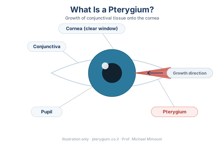
“Surfer’s eye” is the everyday name for the condition doctors call a pterygium. Many people search for the nickname rather than the medical term, so it is worth saying plainly at the outset: these are not two different conditions. They are the same thing — a growth of fibrovascular tissue that begins on the conjunctiva, the membrane covering the white of the eye, and can advance onto the cornea, the transparent window at the front of the eye.
Where the name comes from
The nickname reflects the classic risk profile: long hours outdoors in bright sun, with wind, water glare and sand. But you do not need to have ever surfed. The underlying driver is cumulative exposure to ultraviolet light, so farmers, builders, fishermen, athletes and anyone who spends substantial time outdoors in a sunny climate can develop one.
What it looks like
A pterygium usually appears as a fleshy, wedge- or wing-shaped growth that starts at the inner corner of the eye — the side nearest the nose — and slowly extends toward the pupil. It is often pinkish, and small blood vessels may be visible within it. Early on it may be small and cause no trouble at all; as it advances it becomes more visible and more noticeable.
Symptoms
Common symptoms include dryness, redness, irritation, tearing and the sensation of something in the eye — as though a grain of sand were caught there. Some people have no symptoms at all and notice the growth only in the mirror or during a routine eye examination. Once the growth reaches the cornea it can flatten or distort its curvature, leading to astigmatism and blurred vision.
What causes it
The principal cause is cumulative UV exposure. Wind, dust and dry air contribute as well. Wearing UV-blocking sunglasses and a wide-brimmed hat is among the most effective ways to reduce the risk.
Surfer’s eye and similar findings
Not every growth on the white of the eye is a pterygium. Another common finding is pinguecula — a yellowish growth on the conjunctiva that does not cross onto the cornea. Distinguishing between the two matters, because it changes the treatment recommendation, and it is done by an ophthalmologist.
How it is diagnosed
Diagnosis is relatively straightforward: an ophthalmologist examines the surface of the eye with a slit lamp — a specialised microscope — and assesses the position, size and extent of the growth, and whether it has reached the cornea. In more advanced cases, corneal measurements can be added to assess the effect on vision.
Treatment options
Mild cases are managed conservatively with lubricating and anti-inflammatory drops, which relieve dryness and irritation without removing the growth. Surgery is considered when the pterygium advances toward the centre of the cornea, affects vision, causes persistent discomfort that does not respond to conservative measures, or is cosmetically bothersome.
The current approach is excision combined with a conjunctival autograft secured with tissue glue, usually without sutures. This replaces the abnormal tissue with healthy tissue and substantially lowers the risk of recurrence compared with older methods.
When to seek care
It is worth being examined when the growth changes in size or appearance, when irritation or redness persists, or when there is any change in vision. Early assessment allows the growth to be monitored and treatment planned in good time — before it reaches the central cornea and leaves scarring.
Frequently asked questions
Is surfer’s eye the same as a pterygium?
Do you have to be a surfer to get it?
Is surfer’s eye dangerous?
Does every pterygium need surgery?
Have a question about pterygium? Want a specialist’s opinion?